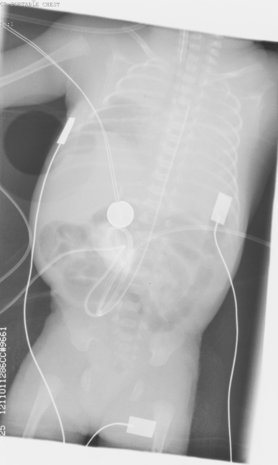
There is consolidation in the left lung and RUL. Ground glass opacification RLZ. Endotracheal tube in situ. It lies a little high in position and ideally should be between T1 and T2. The umbilical artery catheter tip is at T4. Ideally this should be sited at T6-T10. No significant bowel gas abnormality. The appearances are highly suggestive of RDS (hyaline membrane disease).